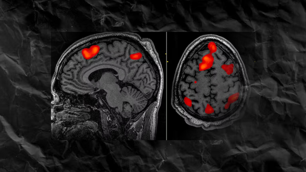
New Findings Challenge Claims of Direct Neural Activity Imaging

A recent study led by researchers at MIT’s McGovern Institute has cast doubt on the validity of a purportedly revolutionary method for imaging brain activity. Published in Science Advances on March 27, 2024, the study challenges the claims made about a magnetic resonance imaging (MRI) technique known as DIANA, which was initially heralded for its potential to directly detect neural activity.
Functional MRI (fMRI) methods conventionally rely on detecting changes in blood flow as an indirect measure of brain activity. However, DIANA promised to revolutionize this approach by claiming to directly detect the electrical signals of neurons, offering unprecedented speed and precision in imaging neuronal activity.
Alan Jasanoff, Associate Investigator at McGovern Institute, explained the significance of such a breakthrough, stating, “If we could look at the whole brain and follow its activity with millisecond precision and know that all the signals that we’re seeing have to do with cellular activity, this would be just wonderful. It could tell us all kinds of things about how the brain works and what goes wrong in disease.”
Initially, DIANA garnered widespread attention within the neuroscience community for its potential to provide insights into brain function. However, subsequent investigation by Jasanoff’s team, led by postdoctoral researcher Valerie Doan Phi Van, revealed a different story.
Replicating the original experiment, Phi Van observed MRI signals in the brain’s sensory cortex in response to sensory stimulation in rats. However, further tests revealed that these signals were not specific to neuronal activity. Surprisingly, the same signals appeared even when the stimulus was removed, replaced instead with a tube of water. This discrepancy suggested that the purported neural activity signals were in fact artifacts of the imaging process itself.
Phi Van’s meticulous examination traced the source of these misleading signals to a trigger embedded within DIANA’s imaging process. This trigger, synchronized with sensory input delivery, created the illusion of neural activity. Removing or altering this trigger eliminated the false signals, confirming their origin as artifacts of the imaging process rather than genuine neuronal activity.
Moreover, the study highlighted challenges in replicating DIANA’s results and found no evidence supporting the cellular changes proposed by its developers as the source of functional MRI signals.
Jasanoff and Phi Van emphasized the importance of their findings for the scientific community, urging caution in adopting new neuroimaging methods without rigorous scrutiny. While acknowledging the ambition of the original study’s authors, they stressed the necessity of maintaining scientific rigor to ensure accurate advancement in the field of neuroscience.
Follow Neuread.com for more insightful content on neuroscience and cutting-edge research in brain imaging techniques

